42 microscope diagram without labels
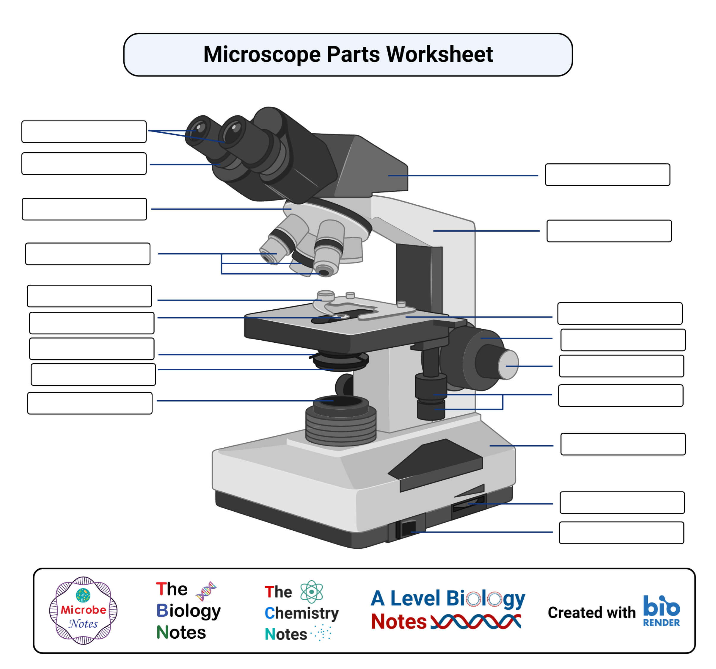
Labeling the Parts of the Microscope | Microscope World Resources Labeling the Parts of the Microscope This activity has been designed for use in homes and schools. Each microscope layout (both blank and the version with answers) are available as PDF downloads. You can view a more in-depth review of each part of the microscope here. Download the Label the Parts of the Microscope PDF printable version here. Label the microscope Diagram | Quizlet Diaphragm. Regulates the amount of light on the specimen. Light Source. Projects light upwards through the diaphragm, the specimen, and the lenses. Arm. supports the body tube. Stage. Supports the slide being viewed. Coarse Adjustment.
How does a Microscope work The optical or light microscope uses visible light transmitted through, refracted around, or reflected from a specimen. Light waves are chaotic; an incandescent light source emits light waves traveling in different paths and of varying wavelengths. Some of the lenses in a microscope bend these light waves into parallel paths, magnify and focus ...

Microscope diagram without labels
Microscope, Microscope Parts, Labeled Diagram, and Functions The Iris Diaphragm is located above the condenser lens and below the microscope stage. The different sized holes in the diaphragm helps to vary the size of the cone and intensity of light that is projected upward into the slide. However, there is no set rule regarding which setting to use for a particular power. Microscope Parts, Function, & Labeled Diagram - slidingmotion Microscope parts labeled diagram gives us all the information about its parts and their position in the microscope. Microscope Parts Labeled Diagram The principle of the Microscope gives you an exact reason to use it. It works on the 3 principles. Magnification Resolving Power Numerical Aperture. Parts of Microscope Head Base Arm Eyepiece Lens A Study of the Microscope and its Functions With a Labeled Diagram ... Here, unlabeled microscope diagrams have been provided for your perusal, which will help you practice and test your understanding of the instrument. Types of Microscopes Depending on the source of illumination, microscopes can be divided into two categories. They are:
Microscope diagram without labels. PDF Label parts of the Microscope: Answers Label parts of the Microscope: Answers Coarse Focus Fine Focus Eyepiece Arm Rack Stop Stage Clip . Created Date: 20150715115425Z ... Fluorescence - Wikipedia Fluorescence is the emission of light by a substance that has absorbed light or other electromagnetic radiation.It is a form of luminescence.In most cases, the emitted light has a longer wavelength, and therefore a lower photon energy, than the absorbed radiation. Label A Microscope Teaching Resources | Teachers Pay Teachers The 13 parts of the microscope: microscope, base, arm, inclination joint, course adjustment, fine adjustment, body tube, ocular lens, revolving nose piece, objectives, stage, stage clips, and iris diaphragm.Includes:13 cards with labels13 cards without labels13 labels1 blackline masterCards with labels are approx. 3¾" x 4", cards without ... ALEX | Alabama Learning Exchange Students will use a Venn diagram to compare lightning and static electricity. Then, students will experiment with static electricity and read nonfiction passages about lightning and lightning rods. Finally, they will apply their learning to construct a model of a lightning rod system that protects a house from a lightning-induced fire.
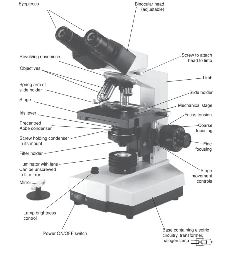
Endomembrane system - Definition and Examples - Biology ... Jun 16, 2022 · Figure 14: Layers of the Epidermis: (left) schematic diagram showing parts and (right) microscope image of the epidermis showing the stratum basale to the stratum corneum. Source: Modified by Maria Victoria Gonzaga of Biology Online, from the works of LumenLearning.com (epidermal layers diagram) and Mikael Häggström et al., CC BY 3.0. Binocular Microscope Anatomy - Parts and Functions with a Labeled Diagram Now, I will discuss the details anatomy of the light compound microscope with the labeled diagram. Why it is called binocular: because it has two ocular lenses or an eyepiece on the head that attaches to the objective lens, this ocular lens magnifies the image produced by the objective lens. Binocular microscope parts and functions Microscope Label Interactive Worksheets & Teaching Resources | TpT 12. $1.89. PDF. Students will complete a timeline of the history of the microscope, label a diagram, and create a pocket foldable with terms and definition cards. The timeline can be completed according to the teacher's directions or like the answer key example. Optional cut & paste images and a QR code are a. 22 Parts Of a Microscope With Their Function And Labeled Diagram The field diaphragm control is located around the lens located in the base. Hinge Screw -This screw fixes the arm to the base and allow for the tilting of the arm. Stage Clips - They hold the slide firmly onto the stage. On/OFF Switch - This switch on the base of the microscope turns the illuminator off and on.
Microscope Parts and Functions First, the purpose of a microscope is to magnify a small object or to magnify the fine details of a larger object in order to examine minute specimens that cannot be seen by the naked eye. Here are the important compound microscope parts... Eyepiece: The lens the viewer looks through to see the specimen. Parts of the Microscope with Labeling (also Free Printouts) A microscope is one of the invaluable tools in the laboratory setting. It is used to observe things that cannot be seen by the naked eye. Table of Contents 1. Eyepiece 2. Body tube/Head 3. Turret/Nose piece 4. Objective lenses 5. Knobs (fine and coarse) 6. Stage and stage clips 7. Aperture 9. Condenser 10. Condenser focus knob 11. Iris diaphragm PDF Parts of a Microscope Printables - Homeschool Creations Label the parts of the microscope. You can use the word bank below to fill in the blanks or cut and paste the words at the bottom. ... without needing to move the microscope ? the head •What is the magnification level on the eyepiece of a microscope?10x (see objective Labelled Diagram of Compound Microscope The below mentioned article provides a labelled diagram of compound microscope. Part # 1. The Stand: The stand is made up of a heavy foot which carries a curved inclinable limb or arm bearing the body tube. The foot is generally horse shoe-shaped structure (Fig. 2) which rests on table top or any other surface on which the microscope in kept.
Microscope Diagram - Free Printable Tests and Worksheets ...
Comparative genomic hybridization - Wikipedia Comparative genomic hybridization (CGH) is a molecular cytogenetic method for analysing copy number variations (CNVs) relative to ploidy level in the DNA of a test sample compared to a reference sample, without the need for culturing cells.

Simple Microscope - Diagram (Parts labelled), Principle ...
Microscope Labeling Activity | Sticky Note Labels - Classful This clear and simple microscope labeling activity is exactly what your students need to learn the parts of a compound microscope! There are five versions of microscope labeling diagrams included: ⭐Sticky Note microscope to label ⭐Microscope to label with a word bank ⭐Microscope to label without a word bank ⭐Plain microscope to label without lines ⭐Version with and without name/date ...

Microscope labeled diagram
Microscope Labeling - The Biology Corner Students label the parts of the microscope in this photo of a basic laboratory light microscope. Can be used for practice or as a quiz. ... The type of microscope used in most science classes is the _____ microscope. 18. You should carry the microscope by the _____ and the _____. 19. The objectives are attached to what part of the microscope ...
Parts of a microscope with functions and labeled diagram
Free Microscope Worksheets for Simple Science Fun for Your ... Parts of a Microscope . The first worksheet labels the different parts of a microscope, including the base, slide holder, and condenser. If you have a microscope, compare and contrast this worksheet to it. Also, your kids can color this microscope diagram in and read the words to each part of the microscope.

How to draw compound of Microscope easily - step by step
Simple Microscope - Parts, Functions, Diagram and Labelling A simple microscope is a device that only has one lens for magnification. It functions the same way as the magnifying glass. Although it is simple in terms of design and function, it is useful I various fields including medicine, jewelry and watchmaking, and agriculture, to name a few. References

Dissecting Stereo Microscope Parts and Functions
Label the microscope — Science Learning Hub In this interactive, you can label the different parts of a microscope. Use this with the Microscope parts activity to help students identify and label the main parts of a microscope and then describe their functions. Drag and drop the text labels onto the microscope diagram.

Compound Microscope Parts – Labeled Diagram and their ...
Parts of Stereo Microscope (Dissecting microscope) – labeled ... Labeled part diagram of a stereo microscope Major structural parts of a stereo microscope. There are three major structural parts of a stereo microscope. The viewing Head includes the upper part of the microscope, which houses the most critical optical components, including the eyepiece, objective lens, and light source of the microscope.
Microscope labelling - Teaching resources
Compound Microscope Parts, Functions, and Labeled Diagram Compound Microscope Definitions for Labels. Eyepiece (ocular lens) with or without Pointer: The part that is looked through at the top of the compound microscope. Eyepieces typically have a magnification between 5x & 30x. Monocular or Binocular Head: Structural support that holds & connects the eyepieces to the objective lenses.
How to Use the Microscope
Label Microscope Diagram - EnchantedLearning.com Using the terms listed below, label the microscope diagram. arm - this attaches the eyepiece and body tube to the base. base - this supports the microscope. body tube - the tube that supports the eyepiece. coarse focus adjustment - a knob that makes large adjustments to the focus. diaphragm - an adjustable opening under the stage, allowing ...

label microscope diagram | Charts | Microscope, Anatomy bones ...
19+ Animal Cell Diagram Without Labels Best A simple diagram of an unspecialised animal cell without labels. The most important structures of plant and animal cells are shown in the diagrams below, which provide a clear illustration of how much these cells have in common. The first is a colored and labeled cell diagram. Cells are covered by a cell membrane and come in many different shapes.

microscope | Types, Parts, History, Diagram, & Facts | Britannica
Label the Microscope Diagram | Download Scientific Diagram - ResearchGate Download scientific diagram | Label the Microscope Diagram from publication: Laboratory Exercises in Microbiology: Discovering the Unseen World through Hands-on Investigation | Microbiology ...

The microscope - Natural Sciences Grade 9 - OpenStax CNX
Diagram of a Compound Microscope - Biology Discussion 1. It is noted first that which objective lens is in use on the microscope. 2. Stage micrometer is positioned in such a way that it is in the field of view. 3. The eyepiece is rotated so that the two scales, the eyepiece or ocular scale and the stage micrometer scale, are parallel. 4.

Compound Light Microscope Labeling Diagram | Quizlet
Simple Squamous Epithelium under a Microscope with a Labeled Diagram ... Simple squamous epithelium under microscope labeled in renal corpuscle The cortex of a kidney consists of renal corpuscles and the convoluted tubule, straight tubules, nephrons, connecting tubules, and collecting ducts. You will find the medullary ray in the medulla of the kidney that comprises straight tubules and collecting ducts.
Microscope Clipart Black And White - Light Microscope Black ...
16 Parts of a Compound Microscope: Diagrams and Video Once you have an understanding of the parts of the microscope it will be much easier to navigate around and begin observing your specimen, which is the fun part! The 16 core parts of a compound microscope are: Head (Body) Arm Base Eyepiece Eyepiece tube Objective lenses Revolving Nosepiece (Turret) Rack stop Coarse adjustment knobs
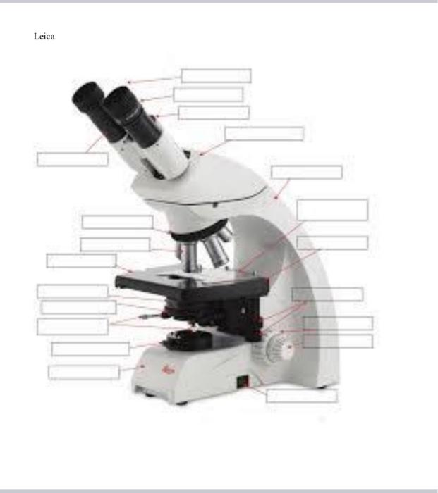
The Compound Light Microscope Label the following parts on ...
Autoclave - Wikipedia A medical autoclave is a device that uses steam to sterilize equipment and other objects. This means that all bacteria, viruses, fungi, and spores are inactivated. However, prions, such as those associated with Creutzfeldt–Jakob disease, and some toxins released by certain bacteria, such as Cereulide, may not be destroyed by autoclaving at the typical 134 °C for three minutes or 121 °C for ...
Solved Nikon Parts of the compound microscope Write the ...
Compound Microscope Parts - Labeled Diagram and their Functions The eyepiece (or ocular lens) is the lens part at the top of a microscope that the viewer looks through. The standard eyepiece has a magnification of 10x. You may exchange with an optional eyepiece ranging from 5x - 30x. [In this figure] The structure inside an eyepiece. The current design of the eyepiece is no longer a single convex lens.

Compound Microscope Parts, Functions, and Labeled Diagram ...
A Study of the Microscope and its Functions With a Labeled Diagram ... Here, unlabeled microscope diagrams have been provided for your perusal, which will help you practice and test your understanding of the instrument. Types of Microscopes Depending on the source of illumination, microscopes can be divided into two categories. They are:
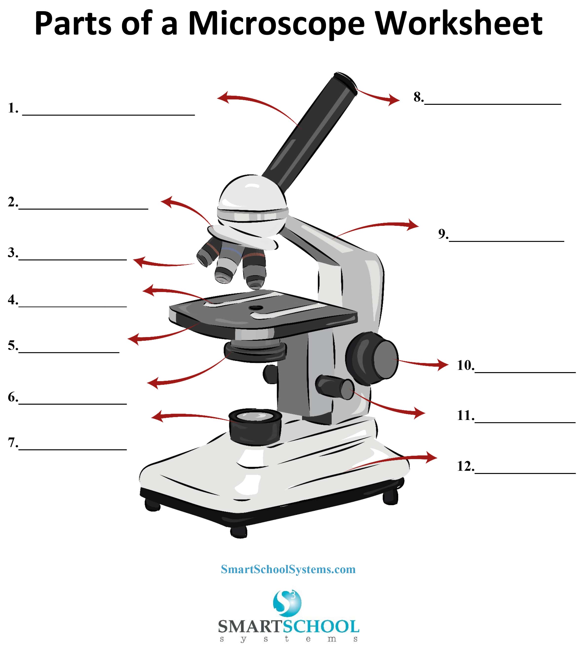
Parts of a Microscope - SmartSchool Systems
Microscope Parts, Function, & Labeled Diagram - slidingmotion Microscope parts labeled diagram gives us all the information about its parts and their position in the microscope. Microscope Parts Labeled Diagram The principle of the Microscope gives you an exact reason to use it. It works on the 3 principles. Magnification Resolving Power Numerical Aperture. Parts of Microscope Head Base Arm Eyepiece Lens
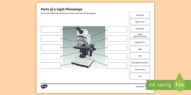
Parts of a Light Microscope Activity | Labeling Task
Microscope, Microscope Parts, Labeled Diagram, and Functions The Iris Diaphragm is located above the condenser lens and below the microscope stage. The different sized holes in the diaphragm helps to vary the size of the cone and intensity of light that is projected upward into the slide. However, there is no set rule regarding which setting to use for a particular power.
National Middle School Standard Microscope, Monocular Head ...
Parts of a Microscope with Their Functions – Microbe Online

This is a common compound microscope Label its parts class 11 ...

SOLVED: Directions: Label the microscope below: Nto: Identify ...

Labeling the Parts of the Microscope | Microscope World Resources

Microscope Labeling

Simple Microscope - Diagram (Parts labelled), Principle ...
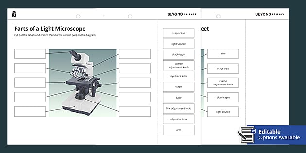
Parts of a Light Microscope Cut and Stick Worksheet - Twinkl

Ms. Chea's Science Class: Microscopes
Microscope Quiz

Parts of Stereo Microscope (Dissecting microscope) – labeled ...
Label a microscope - Teaching resources
Microscope

Compound Microscope – Diagram (Parts labelled), Principle and ...

Lable the microscope worksheet
LAB 1: Scientific Method/Tools of Scientific Inquiry

Microscope, Microscope Parts, Labeled Diagram, and Functions
Compound Microscope - Types, Parts, Diagram, Functions and ...

This is a common compound microscope. Label its parts from A ...
unlabelled diagram of the microscope - Clip Art Library
The Compound Light Microscope Label the following parts on ...
Simple Microscope - Diagram (Parts labelled), Principle ...

Parts of the Microscope with Labeling (also Free Printouts ...
Post a Comment for "42 microscope diagram without labels"